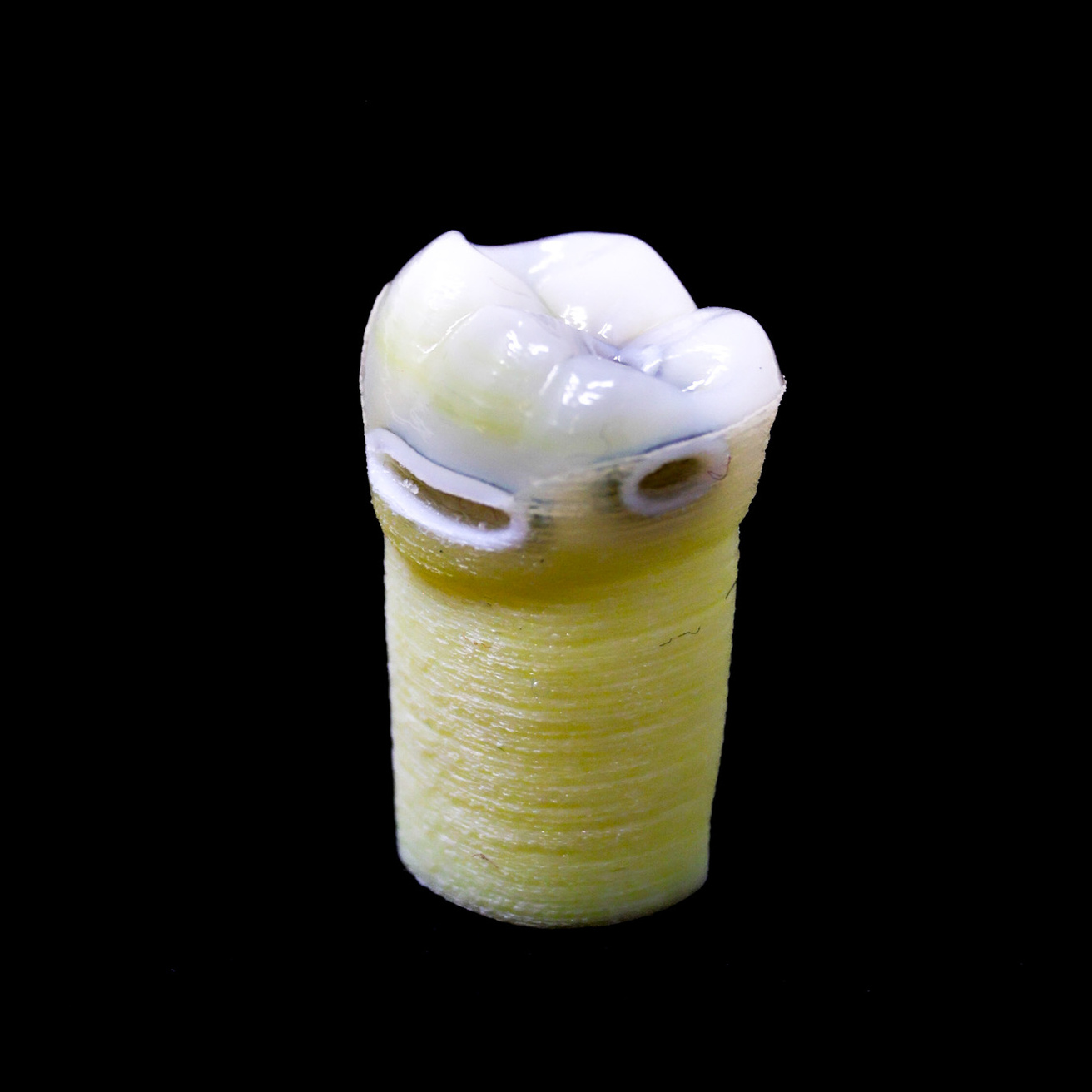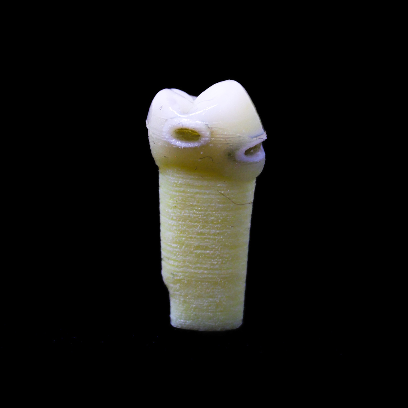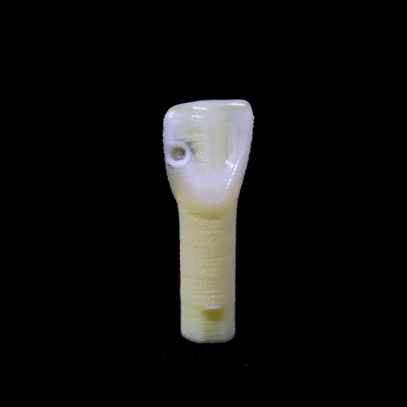
The Educational Caries Teeth are realistic, 3D printed training teeth designed by Dentists at the University of Iowa for educational and training purposes in dentistry, with three different dentin layers that simulate a natural tooth caries lesion. The 3 layers are similar to soft, leathery, and firm dentin. All caries lesions are cavitated with demineralized or chalky enamel visually showing in the lesion margins. All teeth simulated caries lesions ICDAS 5, with different surfaces involved depending on the tooth. The teeth allow students to learn different tactile consistency and texture of dentin layers in caries and practice either selective or complete caries removal.
Each tooth in the pack is identical, including its caries, providing consistency for training and evaluation not feasible with cadaveric teeth.
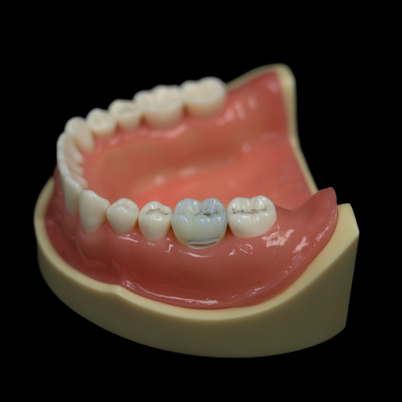
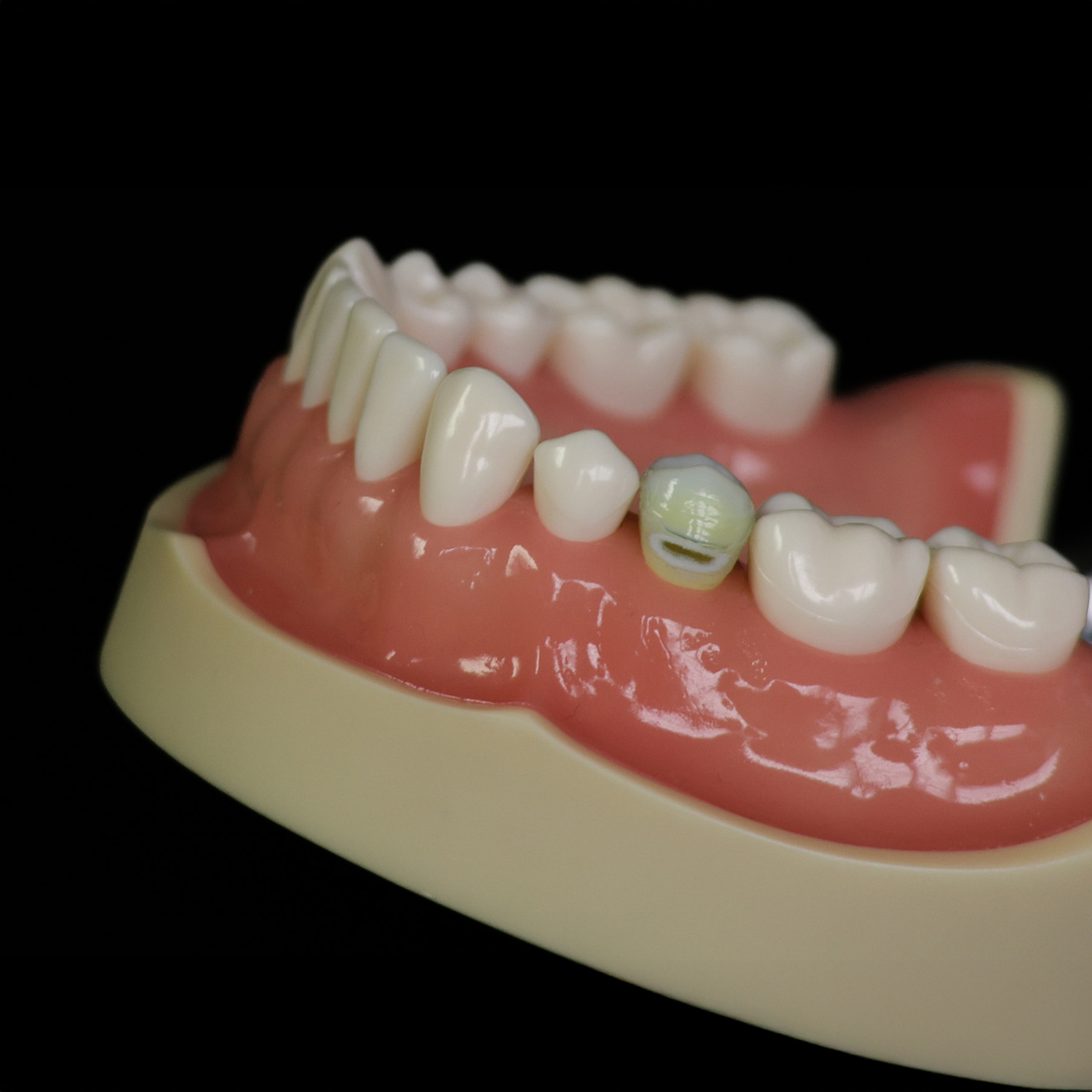
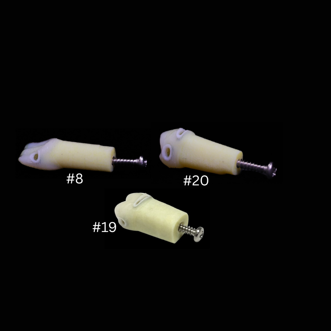
Teeth are numbered using US system
- #8 - one caries lesion involving distal and lingual surfaces
- #14 - two caries lesions (distal, lingual)
- #19 - three caries lesions including one lesion on the occlusal connected to the mesial, one lesion on the distal, and one lesion on the buccal surface
- #20 - three caries lesions including one lesion on the mesial, one lesion on the distal, and one lesion on buccal surface
Key Features
True Multilayer Caries Simulation
Educational Caries Teeth include three distinct dentin layers—soft, leathery, and firm—mirroring real caries progression and giving learners authentic tactile feedback when practicing selective or complete caries removal.
Clinically Relevant ICDAS 5 Lesions
All teeth include cavitated ICDAS 5 lesions with demineralized, chalky enamel visible at the margins, accurately representing moderate‑to‑deep caries. Lesion location varies by tooth number (#8, #14, #19, and #20), allowing students to practice caries removal in clinically common patterns and surfaces.
Standardized and Consistent for Assessment
Each tooth in a pack is identical, ensuring uniformity for teaching, calibration, pre‑clinical assessment, and research—something not possible with extracted teeth.
Supports Skill Development Across Operative Dentistry
The realistic anatomy and layered caries structure help students learn:
How to visually identify carious dentin
How to differentiate dentin hardness by feel
How to perform selective or complete caries removal
How to prepare and restore Class I and Class II cavities with confidence
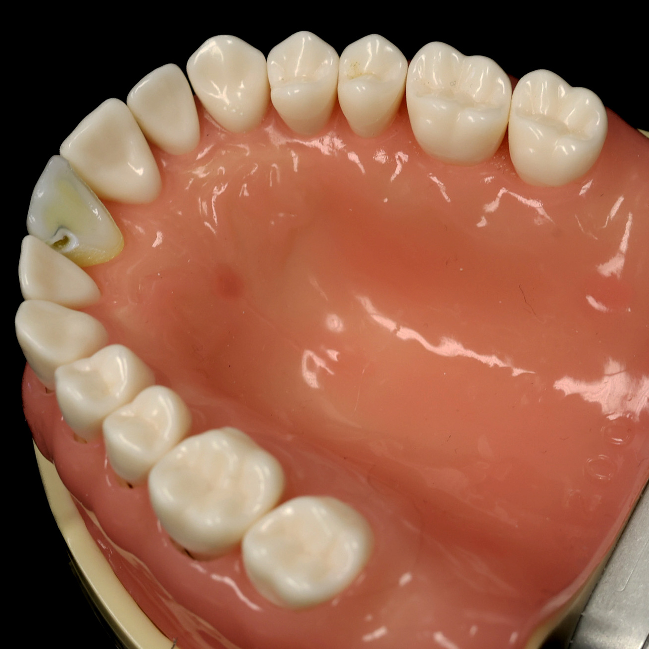
Compatible with Common Typodont Systems
Fits in widely used NISSIN/Kilgore typodont models (PRO2002‑UL‑HD‑DPM‑28 and PRO2002‑UL‑SP‑HM‑28), making integration into existing simulation clinics simple and seamless.
Flexible Purchasing Options
Available individually, in sample packs of 5, or bulk packs of 50, making them ideal for course directors, CE programs, calibration sessions, and research studies.
For training purposes only.